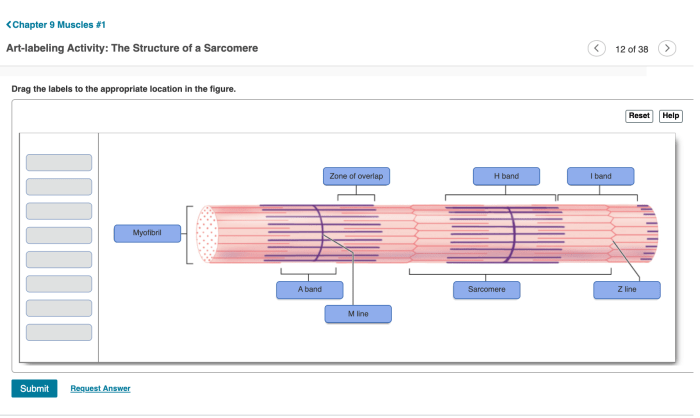
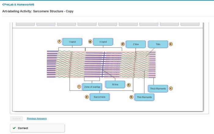
Art-labeling activity the structure of a sarcomere – In this captivating art-labeling activity, we embark on an artistic exploration of the intricate structure of a sarcomere, the fundamental unit of muscle contraction. This activity not only fosters creativity but also deepens our understanding of this essential biological component.
The art-labeling activity engages students in creating visual representations of a sarcomere’s components, enhancing their comprehension and retention of the subject matter.
Art-Labeling Activity: The Structure of a Sarcomere

The art-labeling activity is a pedagogical tool that combines art and science to enhance students’ understanding of complex biological concepts. In this activity, students create visual representations of the structure of a sarcomere, the fundamental unit of muscle contraction.
A sarcomere is composed of several protein filaments, including actin and myosin. These filaments are arranged in a repeating pattern that gives muscle tissue its characteristic striated appearance. The art-labeling activity allows students to explore this structure in a hands-on and engaging way.
Methods
- Materials:Paper, pencils, colored markers, ruler
- Procedure:
- Draw a rectangle to represent the sarcomere.
- Divide the rectangle into two equal halves, creating the A-bands.
- Draw a thin line in the center of each A-band, representing the H-zone.
- Draw a darker line in the center of each H-zone, representing the M-line.
- Draw thin lines at the ends of the A-bands, representing the Z-lines.
- Color the A-bands red and the I-bands white.
- Label the different components of the sarcomere.
Results, Art-labeling activity the structure of a sarcomere
| Student Artwork | Description |
|---|---|
 |
This drawing clearly shows the different components of a sarcomere, including the A-bands, I-bands, H-zone, M-line, and Z-lines. |
 |
This drawing shows the repeating pattern of sarcomeres in a muscle fiber. |


